Sarcoma Kaposi
Introduction
Kaposi sarcoma (Kaposi's sarcoma, KS) was described initially in 1872 by a Hungarian dermatologist, Moritz Kaposi. Kaposi sarcoma is a spindle-cell tumor thought to be derived from endothelial cell lineage. This condition carries a variable clinical course ranging from minimal mucocutaneous disease to extensive organ involvement. Kaposi sarcoma can be primarily categorized into four types: epidemic of AIDS-related, immunocompromised, classic or sporadic, and endemic (African).
- Epidemic AIDS -related Kaposi sarcoma
- This entity occurs in patients with advanced HIV infection and is the most common presentation of Kaposi sarcoma. It is the most common malignancy seen in HIV-infected patients, especially where access to HAART (highly active antiretroviral therapy) is limited.
- In the United States, Kaposi sarcoma serves as an AIDS-defining illness in 2-3% of HIV-infected homosexual men. In the mid 1990s, it was the initial presentation in approximately 15% of homosexual men. In Africa and developing regions, epidemic AIDS-related Kaposi sarcoma is common in heterosexual adults and occurs less often in children. Visceral involvement is widespread.
- AIDS-related Kaposi sarcoma is the most clinically aggressive form of Kaposi sarcoma.
- Seroconversion to human herpesvirus 8 (HHV-8) positivity predates the development of epidemic Kaposi sarcoma by 5-10 years. The interval for development of Kaposi sarcoma is shortened in patients where HIV infection precedes seroconversion to HHV-8 positivity.
- The presence of decreased CD4 counts and increased HIV-1 viral loads are independent prognostic factors in the development of epidemic Kaposi sarcoma. Less than one-sixth of HIV-infected patients have CD4 count of over 500 per microliter. The disease usually develops in HIV infected patients with severe immunodeficiency.
- Cigarette smoking may be protective for Kaposi sarcoma risk in HHV-8 seropositive patients infected with HIV.
- Relative affluence may increase the risk of Kaposi sarcoma in HIV positive patients.
- Immunocompromised Kaposi sarcoma
- This entity can occur following solid-organ transplantation or in patients receiving immunosuppressive therapy. The incidence of Kaposi sarcoma is increased 100-fold in transplant patients However, individuals with congenital immunodeficient states are not at increased risk for developing Kaposi sarcoma.
- This form of Kaposi sarcoma is rare8 but is more common in patients at risk for classic Kaposi sarcoma.
- The average time to development of Kaposi sarcoma following transplantation is 15-30 months. An aggressive course is the rule with visceral involvement being common. However, withdrawal of immunosuppression may cause regression of the disease.10 This is further evidence for the important role that immune suppression may play in the development of Kaposi sarcoma. Immune activation and suppression both play roles to affect the natural history of HHV-8 in a very complex manner.
- Clearly the immunotherapy required to prevent rejection puts patients at greater risk for developing Kaposi sarcoma. One such drug, sirolimus (Rapamune) has been shown to have simultaneous antitumor effect as well as the immunosuppressive effect required to prevent graft rejection.
- In a study of 15 kidney-transplant patients switched from cyclosporine to sirolimus with biopsy-proven Kaposi sarcoma, all cutaneous lesions in all patients disappeared. levels of Flk-1/KDR, phosphorylated Akt, p70S6, and vascular endothelial growth factor (VEGF) were increased in Kaposi sarcoma cells. It appears as though sirolimus has an anti-Kaposi sarcoma mechanism of action independent of its immunosuppressive mechanism.
- No rejection was noted in the patients on this drug. The mechanism may be related to both its antiangiogenesis effect with reduction of VEGF and its Flk-1/KDR receptor on the tumor cells and inhibition of the mTOR pathway by blocking of Akt. mTor can activate various mediators of proliferation and affect control points of translation in the cell cycle. This dual activity may be helpful in providing immunosuppressive therapy without increased risk of Kaposi sarcoma development in patients undergoing organ transplantation.
- Classic (sporadic) Kaposi sarcoma
- This entity typically occurs primarily in elderly men of Mediterranean and Eastern European background. It has a male predominance with a male-to-female ratio of 10-15:1. The age of onset is between 50 and 70 years.
- Classic Kaposi sarcoma usually carries a protracted and indolent course. Common complications include venous stasis and lymphedema. This form of the disease rarely has lymph node, mucous membrane, or visceral involvement.As many as 30% of patients with classic KS subsequently may develop a second malignancy, typically a non-Hodgkin lymphoma.Its occurrence may be due to immune suppression from age, host genetics, history of other neoplasm, and possible concurrent infections such as malaria.Contradictory evidence suggests that immune activation is requisite for the development of classic Kaposi sarcoma. Indeed both hypotheses may be correct, indicating a complicated mechanism of immune dysregulation.
- Infrequent bathing, a history of asthma, and in men, a history of allergy have been linked to classic Kaposi sarcoma among Italian patients.Topical use of steroids is associated with increased risk, either indicating an effect of chronic dermatitis or of the steroids themselves. A finding similar to that seen in epidemic AIDS-related Kaposi sarcoma with regard to the protective effect of cigarette smoking has also been noted.
- Endemic African Kaposi sarcoma
- This entity occurs primarily in men but also in women and children who are HIV seronegative in Africa and may carry an indolent or aggressive course. It was relatively common before the AIDS epidemic. Since the advent of AIDS, it has increased about 20-fold. In the African countries of Malawi, Swaziland, Uganda, Zambia, and Zimbabwe.
- Kaposi sarcoma has become the most prevalent form of cancer in men and the second in incidence among women. It represents about 9% of all cancers seen in Ugandan males. The disease involves lymph nodes more commonly than the classic variant. Factors associated with risk among HHV-8 seropositive patients are not as well characterized.
- The rare wearing of shoes is associated with an increase of endemic Kaposi sarcoma.Relative affluence in Ugandan patients is associated with Kaposi sarcoma, a finding similar to that of Kaposi sarcoma in AIDS patients. Whether this represents local immune suppression due to chronic lymphatic obstruction from fine soil particles may go hand in hand with the association of classic KS with use of topical steroids.
A lymphadenopathic form of Kaposi sarcoma is also seen in Africa, chiefly in HIV seronegative children before the age of puberty. Generalized lymphadenopathy is seen with visceral involvement and carries a poor prognosis with a 100% fatality rate at 3 years.
The common theme of immune dysregulation is associated with all 4 types of Kaposi sarcoma. Diminished responsiveness of cytotoxic T-lymphocytes is associated with Kaposi sarcoma pathogenesis.Restoration of NK cell cytoxic effect may explain regression of Kaposi sarcoma in AIDS patients treated with HAART.There may be immune activation in Kaposi sarcoma as well with a role for inflammatory cytokines such as gamma interferon and the initiation of HHV-8 infected cell proliferation by HIV-tat proteinThis complex interaction of HIV, HHV-8, environmental factors, and the immune system requires further investigation to attempt to decipher the true pathogenesis of Kaposi sarcoma.
Finally, there have been several reports of epidemic Kaposi sarcoma in homosexual men without a shred of evidence of HIV infection. These men usually have CD4 counts of greater than 300/mL. This has been seen in primarily an indolent cutaneous form. There may be other factors than HIV at play in this case.
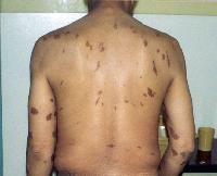
Epidemic Kaposi sarcoma (KS). Large violaceous truncal nodules with typical linear and symmetric distribution pattern.
Pathophysiology
Kaposi sarcoma (Kaposi's sarcoma, KS) is caused by an excessive proliferation of spindle cells thought to have an endothelial cell origin. Despite their heterogeneity, the tumors are predominantly composed of Kaposi sarcoma human herpesvirus (KSHV) genomic material with immunohistochemical markers of both lymphoid, spindle, and endothelial cells.1 Although the origin is still unknown, there is increased endothelial factor VIIIa antigen, spindle cell markers such as smooth muscle alpha-actin, and macrophage markers such as PAM-1, CD68, and CD14 expressed by these spindle cells.27 This would suggest a pluripotent mesenchymal progenitor. The spindle cells proliferate in a background of reticular fibers, collagen and mononuclear cells including macrophages, lymphocytes and plasma cells. They tend to be vascular involving in the either the reticular dermis (patch stage) or the entire thickness of the dermis (plaque or nodular stage).
Molecular studies previously suggested that Kaposi sarcoma originates from a single cell clone rather than a multifocal origin. However, more recent data in a study of 98 Kaposi sarcoma patients with primarily cutaneous disease analyzed by molecular diagnostic techniques comparing viral HHV8 DNA of the tumors showed that nearly 80% of the tumors arose independently from multiple cells.The conclusion reached was that few Kaposi sarcoma tumors originate from a single cell and that Kaposi sarcoma may not be metastatic in its advanced form but multifocal and independent occurring at multiple sites. These data are primarily applicable to initially less aggressive cutaneous Kaposi sarcoma. They may not apply for de novo more aggressive visceral Kaposi sarcoma, which may be less likely to respond to therapy.
Human herpesvirus 8 (HHV-8) genomic sequences have been identified by polymerase chain reaction in more than 90% of all types of Kaposi sarcoma lesions (including epidemic and endemic forms), suggesting a causative role for this DNA virus. The current working hypothesis is that HHV-8 must be present for the disease to develop. It is transmitted in saliva. Blood-borne transmission has yet to be proved. HIV significantly increases the risk of immune suppression. These viral sequences additionally have been associated with body cavity-based lymphomas, Castleman disease, and leiomyosarcomas that occur in individuals infected with HIV. Factors that are thought to contribute to the development of Kaposi sarcoma in individuals infected with HHV-8 and HIV include an abnormal cytokine milieu associated with HIV infection with angiogenic cytokines—IL-1 beta, basic fibroblast growth factor (bFGF), acidic fibroblast growth factor, endothelial growth factor, and vascular endothelialgrowthfactor.
Other cytokines include IL-6, granulocyte-monocyte colony stimulating factor (GM-CSF), transforming growth factor beta (TGF-beta), tumor necrosis factor (TNF), and platelet-derived growth factor alpha (PDGF-alpha from interstitial and mononuclear cells. Oncostatin M, IL-1, IL-6, fibroblast growth factor, tumor necrosis factor (TNF), and the HIV-tat protein all of which originate from HIV-infected T cells act as costimulants for Kaposi sarcoma cells. Indeed the later TAT gene may be a key component responsible for conversion of the Kaposi sarcoma cell to a malignant phenotype. A specific viral gene, ORF74, encodes for a G-protein coupled receptor that causes production of VEGF and other angiogenic mediators.
Thus, it appears that Kaposi sarcoma may be caused by HHV-8 (KSHV) with stimulation by autocrine and paracrine growth factors secreted by the spindle cells themselves as well as the supporting network of mononuclear and endothelial cells. Coinfection with HIV may create a more aggressive course, which is mitigated by effective antiretroviral therapies. Indeed, the risk of Kaposi sarcoma development is amplified 500-10,000 times in patients coinfected with KSHV and HIV. The use of technologies such as comprehensive genetic profiling with gene expression arrays may further elucidate the very complicated viral gene-host interaction and facilitate identification of molecular targets for both prevention and treatment.
Frequency United States
Before the AIDS epidemic, Kaposi sarcoma (Kaposi's sarcoma, KS) was rare. Between 1975 and 1980, the incidence in males aged 20-54 years was 19 cases according to Surveillance, Epidemiology, and End Results (SEER) data (0.1 cases per 100,000). In 1981, an aggressive form of Kaposi sarcoma began to appear among homosexual men as one of the harbingers of the AIDS epidemic.At the beginning of the AIDS epidemic, just before 1980, 40-50% of homosexual men with AIDS developed Kaposi sarcoma.
The rate in all SEER areas increased from in the late 1970s to 17.5 per 100,000 in the late 1980s and then decreased to 2.2 per 100,000 as of 1999-2000.In the United States, the risk of Kaposi sarcoma among sexually active homosexual men is much greater than among others infected with HIV.The incidence of Kaposi sarcoma reached its zenith in 1989 among white men aged 20-54 years when it was the most common AIDS-associated neoplasm. Its incidence has dramatically declined since then. In the mid 1990s, approximately 1 in 4 homosexual men contracted the disease. This number has decreased precipitously with the advent of safer sexual practices in the early 1990s and accelerated with the introduction of HAART in the mid 1990s due to improved immune reconstitution. The dramatic decrease supports the hypothesis of the need for severe immunosuppression for the presumed sexual transmission of an infection agent such as Kaposi sarcoma-associated herpesvirus/human herpesvirus 8 (KSHV/HHV-8).
Men in the San Francisco area manifested the most profound decrease from 7.9 to 1.6 cases per 100,000.The incidence in other HIV risk groups initially was 10% in IV drug abusers, 4% in hemophiliacs, and 3% in children with AIDS.It has decreased in these groups as well to a relatively steady rate of 2%, which is now the same rate for homosexual men. The disease may be contracted by other groups with HIV, such as women and heterosexual men, through unprotected sex. Overall, there has been a historic tendency to underreport Kaposi sarcoma in the AIDS population.
he other major group in the United States in whom Kaposi sarcoma occurs is the posttransplant population, in whom the incidence is about 1 in 200. Approximately 2,500 cases of Kaposi sarcoma occur yearly in the United States at the present time. The incidence among African-American men peaked somewhat later than in white males, in 1991-1999. It was first noted in Hispanic men in 1992 when its incidence was transiently higher than in African-Americans or whites. The dramatic drop in incidence has been seen in all major ethnic groups from 1992-2001, with stabilization since then. Now the highest rate is seen in African-American males with a rate of 3 per 100,000 as opposed to rates less than 3 per 100,000 in decreasing incidence for Hispanics, whites, and Asians/Pacific Islanders respectively.
As noted above, the incidence and severity of Kaposi sarcoma has lessened following the introduction of HAART. This reduction has been attributable to restoration of the immune system caused by these drugs. The regression occurs in parallel with increases in CD4 counts usually within no more than 9-12 months. Conversely, progression occurs with increasing viral load, low CD4 counts, and opportunistic infection (OI).Maurer et al have, however, reported a cluster of cutaneous, refractory HIV-associated Kaposi sarcoma in patients in the San Francisco area with CD4 count above 300 cells/mL and suppressed viral loads below 300 copies for at least 2 years. All patients presented between November, 2004 and November, 2006 and were being treated with one protease inhibitor or non-nucleoside RTI. There was no history of opportunistic infection. All the courses were indolent. The hypothesis generated to explain this is that there are a larger number of aging patients infected with both HIV and HHV8.
Also, the specific HAART regimen may be important as the drugs may also act as antitumor or antiangiogenic agents. This phenomenon may not be as rare as previously thought and will require greater monitoring in the future with attention to age, duration of HIV infection, HHV8 viral load, and patterns of viral gene expression.
International
Prior to the advent of HIV, Kaposi sarcoma was common in central Africa and prevalent in Mediterranean countries and the Middle East. It rarely occurred elsewhere. In Africa, the incidence of Kaposi sarcoma is very high at 37.7 per 100,000 in men and 20.5 per 100,000 in women. In Europe, the highest rates of classic Kaposi sarcoma are in Sicily (Ragusa, 30.1 cases per million in men/5.4 cases per million in women) and Sardinia (24.3 cases per million in men/7.7 cases per million in women).
See related CME at Global Burden of Sexually Transmitted Infections (Slides With Transcripts).
Mortality/Morbidity
AIDS-related Kaposi sarcoma, unlike other forms of Kaposi sarcoma, tends to have an aggressive clinical course. Morbidity may occur from extensive cutaneous, mucosal, or visceral involvement. In patients receiving HAART, the disease often has a more indolent clinical course or may regress spontaneously. The most common causes of morbidity include cosmetically disfiguring cutaneous lesions, lymphedema, gastrointestinal involvement, or pulmonary involvement (see History and Physical). Pulmonary involvement is the most common cause of mortality with uncontrolled pulmonary hemorrhage.
Race
- In Africa and developing regions, epidemic AIDS-related Kaposi sarcoma is common in heterosexual adults and occurs less often in children.
- Classic Kaposi sarcoma typically occurs in elderly men of Mediterranean and Eastern European background.
- Endemic African Kaposi sarcoma occurs in HIV seronegative men in Africa.
Sex
- AIDS-related Kaposi sarcoma: In the United States, this condition occurs primarily in homosexual males, bisexual men, and in the female sexual partners of bisexual men. African Kaposi sarcoma occurs in heterosexual men and women with equal frequency. Classic Kaposi sarcoma occurs primarily in males, with a male-to-female ratio of 10-15:1.
Age
- AIDS-related Kaposi sarcoma generally occurs in young to middle aged adults aged 20-54 years.
- Classic Kaposi sarcoma typically occurs in patients aged 50-70 years.
- African Kaposi sarcoma occurs in people of a younger age (35-40 y).
Clinical
HistoryAIDS-related Kaposi sarcoma (Kaposi's sarcoma, KS) carries a variable clinical course ranging from minimal mucocutaneous disease to widespread organ involvement. The lesions may involve the skin, oral mucosa, lymph nodes, and visceral organs. Most patients present with cutaneous disease. Visceral disease may occasionally precede cutaneous manifestations. Lesions have been reported in autopsy series involving virtually every organ.
- Cutaneous lesions occur in virtually all patients.
- Multiple skin lesions - See Physical for description.
- Usually multicentric in a continuum of development
- Tumor-associated lymphedema - Typically manifested by lower extremity or facial involvement, thought to occur secondary to obstruction of lymphatic channels
- Pain associated with ambulation - Due to lesions involving the soles of the feet
- Gastrointestinal lesions can occur anywhere within the gastrointestinal tract.
- Lesions are often asymptomatic and clinically indolent.
- Gastrointestinal disease is usually an indicator of more advanced HIV infection.
- Symptoms include the following:
- Odynophagia, dysphagia
- Nausea, vomiting, abdominal pain
- Hematemesis, hematochezia, melena
- Bowel obstruction
- Pulmonary involvement may be difficult to distinguish from opportunistic infections.
- Symptoms include the following:
- Cough
- Dyspnea
- Hemoptysis
- Chest pain
- Symptoms include the following:
- Pulmonary lesions may be an asymptomatic radiographic finding.
- Pleural effusions are often exudative and bloody.
Physical
- Cutaneous lesions may occur at any location but typically are concentrated on the lower extremities and the head and neck region.
- Lesions may have macular, papular, nodular, or plaquelike appearances.
- Nearly all lesions are palpable and nonpruritic.
- Lesions may range in size from several millimeters to several centimeters in diameter.
- Lesions may assume a brown, pink, red, or violaceous color and may be difficult to distinguish in dark-skinned individuals.
- Lesions may be discrete or confluent and typically appear in a linear, symmetric distribution, following Langer lines.
- Mucous membrane involvement is common (palate, gingiva, conjunctiva). Ulcerated or bulky tumor involvement may interfere with speech or mastication.
Causes
- Coinfection with HIV and HHV-8 (Kaposi sarcoma-associated herpesvirus [KSHV])
- Iatrogenic immunosuppression (including corticosteroids)
- Elevated degree of expression of numerous cytokines and angiogenic growth factors, including TNF-alpha, IL-6, bFGF, HIV-tat protein, and oncostatin M
Free Consultation
Conditions